Quantitative analysis of imaging data
Introduction
Quantitative imaging (QI) is becoming more widely used in modern radiology, aiding in the clinical evaluation of a wide range of patients and giving biomarkers for various illnesses. QI is frequently used to help with patient diagnosis or prognosis, therapy selection, and therapy response monitoring. Because most radiologists will likely utilise specific QI tools to fulfil their referring physicians’ patient care requirements, all radiologists must understand the benefits and limits of QI.
Digital pictures are used to offer data and information in quantitative image analysis. Due to the vast quantity of information created and gathered, this is done with computer technology to detect patterns, construct maps, and analyse signals inside images that cannot be done with the human eye. Quantitative image analysis may be done in various ways, including medical scanning, object detection, and three-dimensional modelling. Individuals with computer engineering, computer vision, and image analysis understand how to utilise technology to evaluate data conduct the procedures. Quantitative image analysis is used because certain types of pictures are difficult to evaluate without technology assistance.
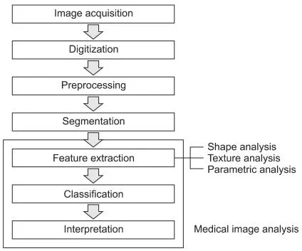
For example, in some medical areas, pictures can be utilised to discover information about what is going on within a person’s body that would usually only be discovered through a surgical operation. Quantitative image analysis is used in security to identify people’s faces and aid in the elimination of security hazards. Several other uses of quantitative image analysis can be helpful in various circumstances and for locating various sorts of data. Medical image analysis is usually carried out systematically, including phases for both quantitative measurements and abstract interpretation of biological pictures (Figure 1).
Figure 1. Medical image analysis procedure [1].
The data obtained from pictures is evaluated using advanced statistical and modelling approaches depending on the final purpose. In some cases, a three-dimensional model is required to give information that would otherwise be difficult, expensive, and time-consuming to get. In other cases, graphs, diagrams, and other quantitative information that might give insight into current patterns are more valuable. Other approaches include looking at the spatial intensity of pixels in an image to analyse incredibly tiny pictures that contain much information. Depending on the goal of the study, information is obtained through quantitative image analysis using methods such as wavelengths of light, cross-sections of materials, and video. After the pictures are acquired, computers are used to convert the data into binary code, which may then be examined further. Because the computer software is so powerful and technological, images may be readily altered, edited, or transformed.
Image analysis is done by statisticians, computer scientists, and engineers in a variety of areas. Individuals who work in this field typically have postgraduate degrees to comprehend the complex computer programming and statistical approaches required to make sense of the massive amounts of data. Quantitative image analysis is a discipline that is continuously changing and growing as new technology is created.
References
[1] Kim, T. Y., Son, J., & Kim, K. G. (2011). The recent progress in quantitative medical image analysis for computer aided diagnosis systems. Healthcare informatics research, 17(3), 143–149. https://doi.org/10.4258/hir.2011.17.3.143 [2] Rosenkrantz, A. B., Mendiratta-Lala, M., Bartholmai, B. J., Ganeshan, D., Abramson, R. G., Burton, K. R., Yu, J. P., Scalzetti, E. M., Yankeelov, T. E., Subramaniam, R. M., & Lenchik, L. (2015). Clinical utility of quantitative imaging. Academic radiology, 22(1), 33–49. https://doi.org/10.1016/j.acra.2014.08.011


